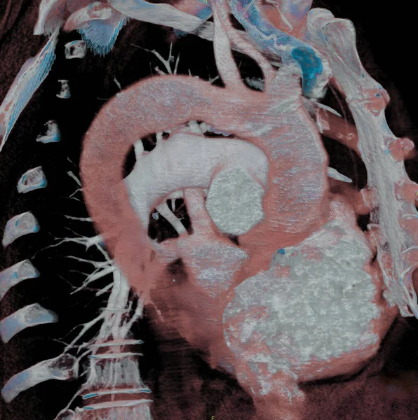
Scan Coronary Bypasses — Stok fotoğraf

3D CT scan of coronary bypasses. — Stok fotoğrafçılık, telifsiz resim

3D CT scan of coronary bypasses.

 — imagepointfr - Fotoğraf

Aynı seride:

RV-PA tube (right ventricle-pulmonary artery). 20-day-old infant born without central pulmonary artery, after operation to place tube between the right ventricle and the right and left pulmonary arteries. 3D CT scan.
Tube Right Ventricle Pulmonary Artery Day Old Infant Born Central — Stok fotoğraf
Anomalous left coronary artery, to the right of the aorta, passes in front of the pulmonary artery. This rare congenital anomaly is, nevertheless, not dangerous since the pulmonary artery does not restrict the coronary artery. 3D CT scan.
Anomalous Left Coronary Artery Right Aorta Passes Front Pulmonary Artery — Stok fotoğraf
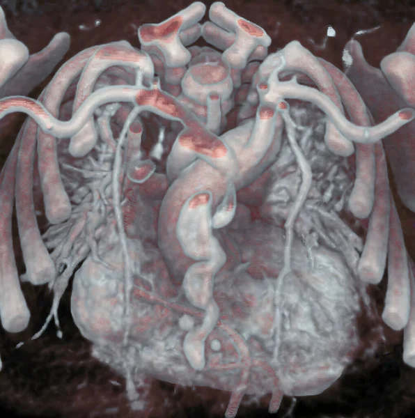
Thorax of newborn : Mediastinum, heart, aorta, and mammary arteries are visible. 3D CT scan.
Thorax Newborn Mediastinum Heart Aorta Mammary Arteries Visible Scan — Stok fotoğraf
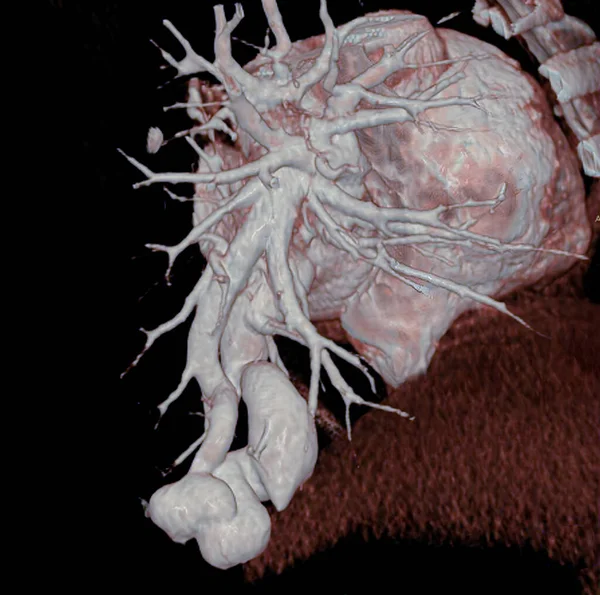
Pulmonary arteriovenous fistula is a congenital anomaly consisting of a direct communication between the pulmonary arterial and venous systems. Complications include intra-pulmonary hemorrhaging (hemoptysis). 3D CT scan.
Pulmonary Arteriovenous Fistula Congenital Anomaly Consisting Direct Communication Pulmonary Arterial — Stok fotoğraf
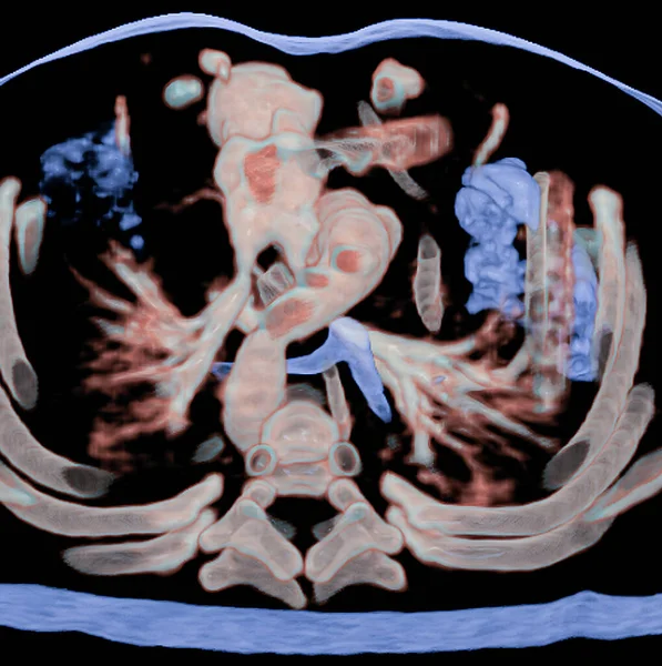
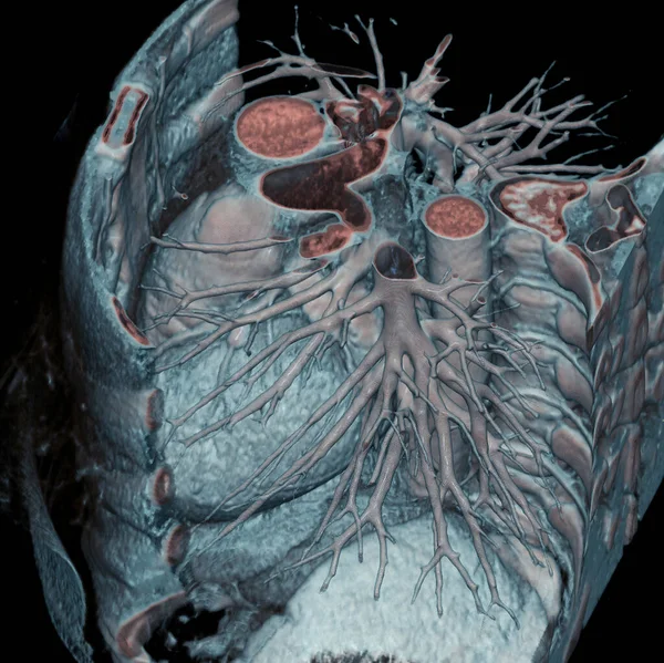
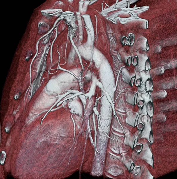
3D CT scan of thorax: lungs, aorta, and pulmonary arteries are visible.
Scan Thorax Lungs Aorta Pulmonary Arteries Visible — Stok fotoğraf
Thorax: 3D CT scan.
Thorax Scan — Stok fotoğraf
Renal artery stenosis (RAS) Extreme stricture (stenosis) of the left renal artery which may be the cause of severe renal-vascular arterial hypertension, 3D CT scan.
Renal Artery Stenosis Ras Extreme Stricture Stenosis Left Renal Artery — Stok fotoğraf
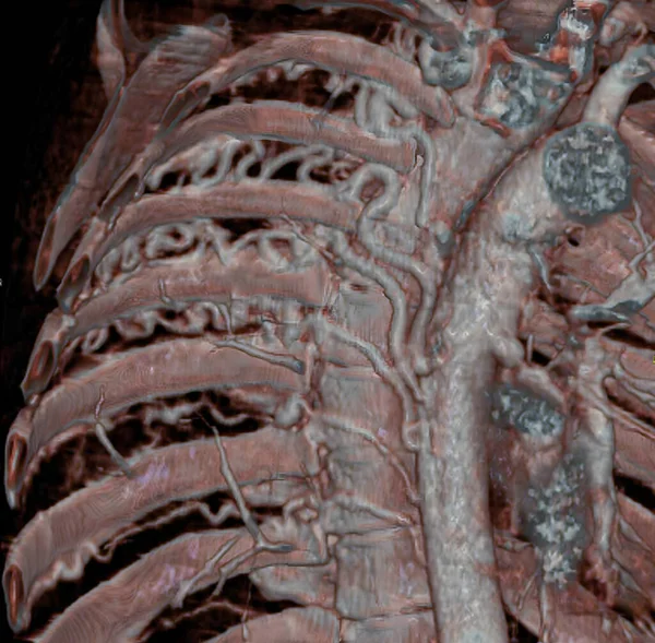
Large intercostal arteries, visible in a CT scan, developed to compensate pulmonary blood flow due to a congenital heart defect characterized by atresia of pulmonary arteries (small or absent pulmonary arteries). 3D CT scan.
Large Intercostal Arteries Visible Scan Developed Compensate Pulmonary Blood Flow — Stok fotoğraf
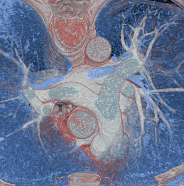
Thorax: Heart, aorta, and pulmonary artery are visible, 3D CT scan.
Thorax Heart Aorta Pulmonary Artery Visible Scan — Stok fotoğraf
Pulmonary artery (PA) aneurysm. Pulmonary aneurysm is associated with pulmonary artery dilation, with a risk of intra-pulmonary hemorrhaging and rupture. 3D CT scan.
Pulmonary Artery Aneurysm Pulmonary Aneurysm Associated Pulmonary Artery Dilation Risk — Stok fotoğraf
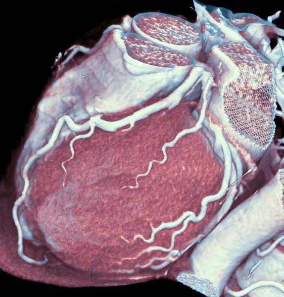
3D CT scan of normal coronary arteries.
Scan Normal Coronary Arteries — Stok fotoğraf
Thorax, 3D CT scan.
Thorax Scan — Stok fotoğraf
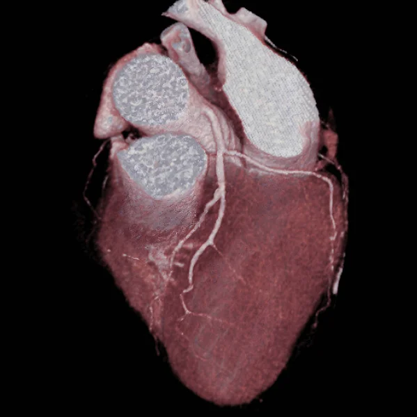
Heart and right coronary artery, 3D CT scan.
Heart Right Coronary Artery Scan — Stok fotoğraf
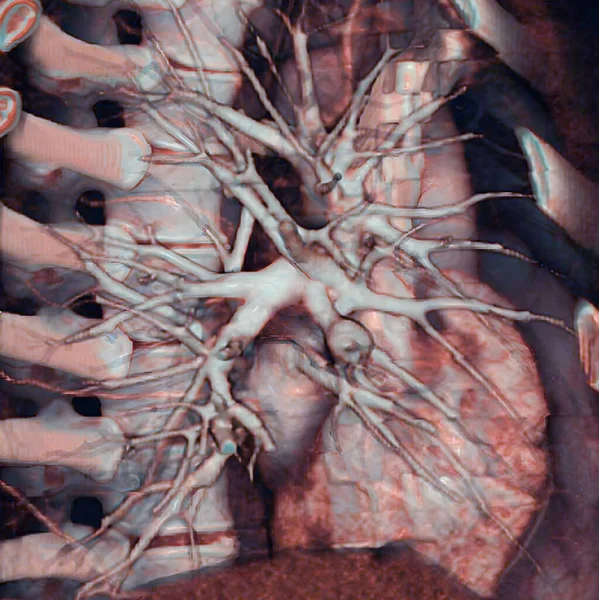
Stenosis of the pulmonary arteries. The 3D CT scan shows few pulmonary arteries. Some are occluded due to a pulmonary embolism.
Stenosis Pulmonary Arteries Scan Shows Few Pulmonary Arteries Some Occluded — Stok fotoğraf
Heart and coronary arteries, 3D CT scan.
Heart Coronary Arteries Scan — Stok fotoğraf
Pelvis and iliac arteries are visible, as well as a stent in the gluteal artery. 3D CT scan.
Pelvis Iliac Arteries Visible Well Stent Gluteal Artery Scan — Stok fotoğraf

Kullanım Bilgisi

Bu telifsiz fotoğrafı " 3D CT scan of coronary bypasses. " Standart veya Genişletilmiş Lisansa göre kişisel ve ticari amaçlarla kullanabilirsiniz. Standart Lisans, reklam, kullanıcı arayüzü tasarımları ve ürün paketleme dahil olmak üzere çoğu kullanım durumunu kapsar ve 500.000'e kadar basılı kopyaya izin verir. Genişletilmiş Lisans, Sınırsız baskı hakkıyla Standart Lisans kapsamındaki tüm kullanım durumlarına izin verir ve indirilen hazır görüntüleri ticari mal, ürün yeniden satışı veya ücretsiz dağıtım için kullanmanıza izin verir.

Bu stok fotoğrafı satın alabilir ve 3020x3171 s'ye kadar yüksek çözünürlükte indirebilirsiniz. Yükleme Tarihi: 11 Ağu 2022