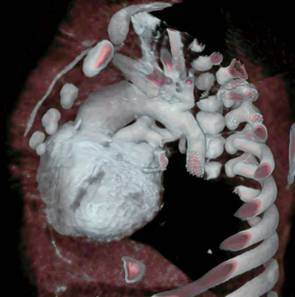
Cervical Vertebrae Scan — Stok fotoğraf

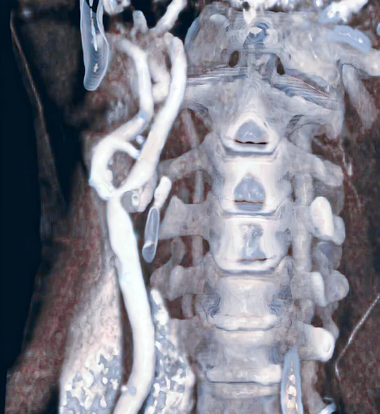
Cervical vertebrae, 3D CT scan. — Stok fotoğrafçılık, telifsiz resim

Cervical vertebrae, 3D CT scan.

 — imagepointfr - Fotoğraf

Aynı seride:

3D CT scan of stenosed carotid artery, responsable for stroke.
Scan Stenosed Carotid Artery Responsable Stroke — Stok fotoğraf
Aortic coarctation of newborn, before surgery, aortic coarctation is a narrowing of the aorta, often considerable and sometimes complete, most commonly just past the point where the aorta and the subclavian artery meet. Aortic coarctation causes low
Aortic Coarctation Newborn Surgery Aortic Coarctation Narrowing Aorta Often Considerable — Stok fotoğraf
Real-time 3D reconstructions from patient images allow personalized 3D printing.
Real Time Reconstructions Patient Images Allow Personalized Printing — Stok fotoğraf
Aortic coarctation is a narrowing of the aorta, often considerable and sometimes complete, most commonly just past the point where the aorta and the subclavian artery meet. Aortic coarctation causes low blood flow, often leading to arterial hypertens
Aortic Coarctation Narrowing Aorta Often Considerable Sometimes Complete Most Commonly — Stok fotoğraf
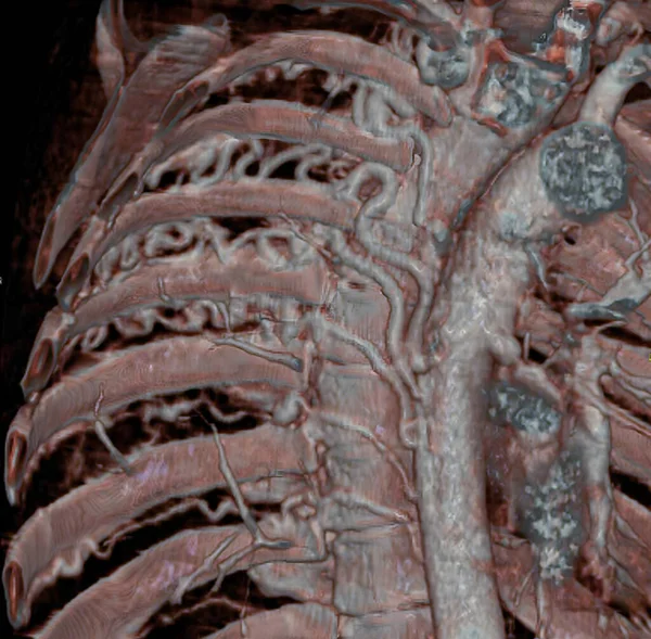
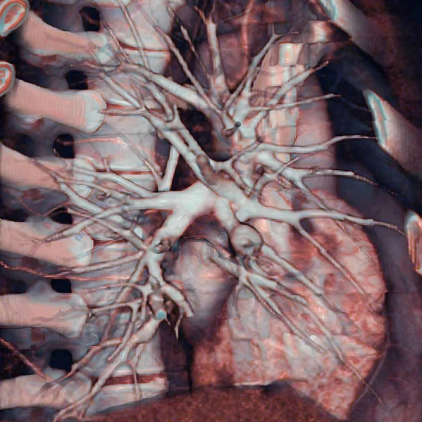
Large intercostal arteries, visible in a CT scan, developed to compensate pulmonary blood flow due to a congenital heart defect characterized by atresia of pulmonary arteries (small or absent pulmonary arteries). 3D CT scan.
Large Intercostal Arteries Visible Scan Developed Compensate Pulmonary Blood Flow — Stok fotoğraf
3D CT scan of coronary arteries and aorta of heart.
Scan Coronary Arteries Aorta Heart — Stok fotoğraf
Thorax: 3D CT scan.
Thorax Scan — Stok fotoğraf
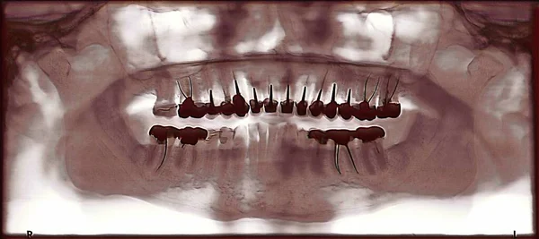
Dental panoramic of a 70 year old person with multiple crowns.
Dental Panoramic Year Old Person Multiple Crowns — Stok fotoğraf
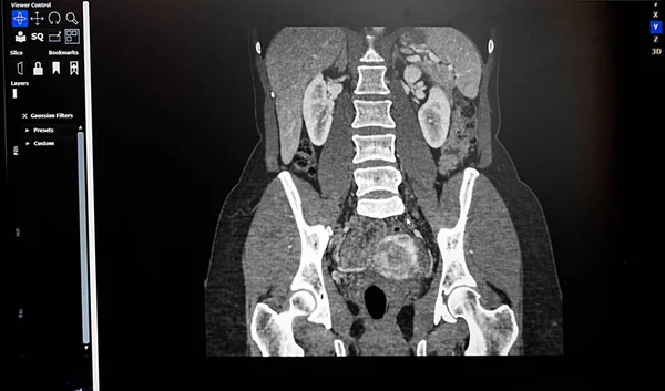
Pelvis and iliac arteries are visible, as well as a stent in the gluteal artery. 3D CT scan.
Pelvis Iliac Arteries Visible Well Stent Gluteal Artery Scan — Stok fotoğraf
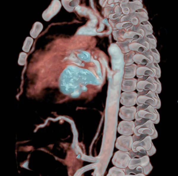
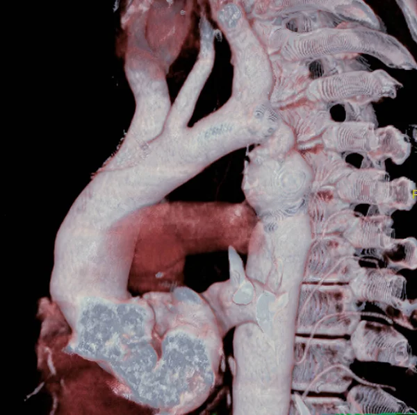
Aortic coarctation. Here, a considerable narrowing of the aorta is visible, just past the point where the aorta and the subclavian artery meet, causing arterial hypertension. 3D CT scan.
Aortic Coarctation Here Considerable Narrowing Aorta Visible Just Point Aorta — Stok fotoğraf
Pulmonary artery (PA) aneurysm. Pulmonary aneurysm is associated with pulmonary artery dilation, with a risk of intra-pulmonary hemorrhaging and rupture. 3D CT scan.
Pulmonary Artery Aneurysm Pulmonary Aneurysm Associated Pulmonary Artery Dilation Risk — Stok fotoğraf
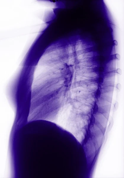
Toraks grafisi
Toraks grafisi — Stok fotoğraf
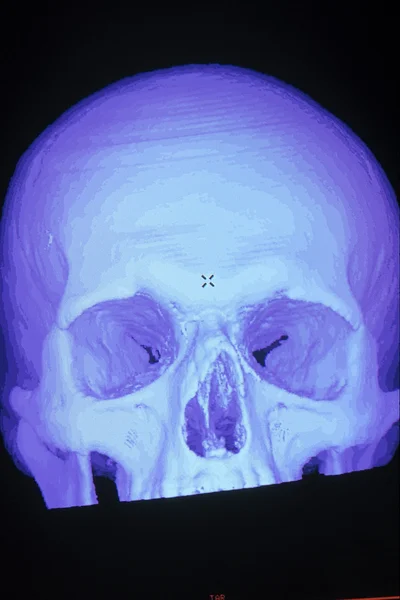
Kafatası siyah arkaplan
Siyah kafatası — Stok fotoğraf
The arterial canal carries blood from the pulmonary artery to the aorta during fetal life and normally closes within hours after birth. In certain cases, however, the canal remains open increasing blood flow into the pulmonary artery, causing increas
Arterial Canal Carries Blood Pulmonary Artery Aorta Fetal Life Normally — Stok fotoğraf
3D CT scan of coronary bypasses.
Scan Coronary Bypasses — Stok fotoğraf
X-ışını resim insan Toraks
Insan toraksı — Stok fotoğraf

Kullanım Bilgisi

Bu telifsiz fotoğrafı " Cervical vertebrae, 3D CT scan. " Standart veya Genişletilmiş Lisansa göre kişisel ve ticari amaçlarla kullanabilirsiniz. Standart Lisans, reklam, kullanıcı arayüzü tasarımları ve ürün paketleme dahil olmak üzere çoğu kullanım durumunu kapsar ve 500.000'e kadar basılı kopyaya izin verir. Genişletilmiş Lisans, Sınırsız baskı hakkıyla Standart Lisans kapsamındaki tüm kullanım durumlarına izin verir ve indirilen hazır görüntüleri ticari mal, ürün yeniden satışı veya ücretsiz dağıtım için kullanmanıza izin verir.

Bu stok fotoğrafı satın alabilir ve 3774x5850 s'ye kadar yüksek çözünürlükte indirebilirsiniz. Yükleme Tarihi: 11 Ağu 2022